Why Surgeons MUST Verify With AI Before Operating
Missing tools cause dangerous mid-surgery delays. Discover how we built an automated vision system using YOLO 11 to precisely segment and track 11 distinct surgical instruments in real-time 4K video, guaranteeing a perfect tray setup before operations even begin.
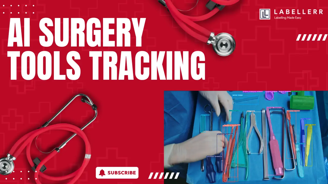
Surgical tools are present in every operating room. You can find scalpels, forceps, and curettes on every surgical tray. In many surgeries, these tools must be perfectly tracked to ensure patient safety. Doing this by hand can take a lot of time and effort. In some high-stress situations, manual tracking can even lead to dangerous mistakes.
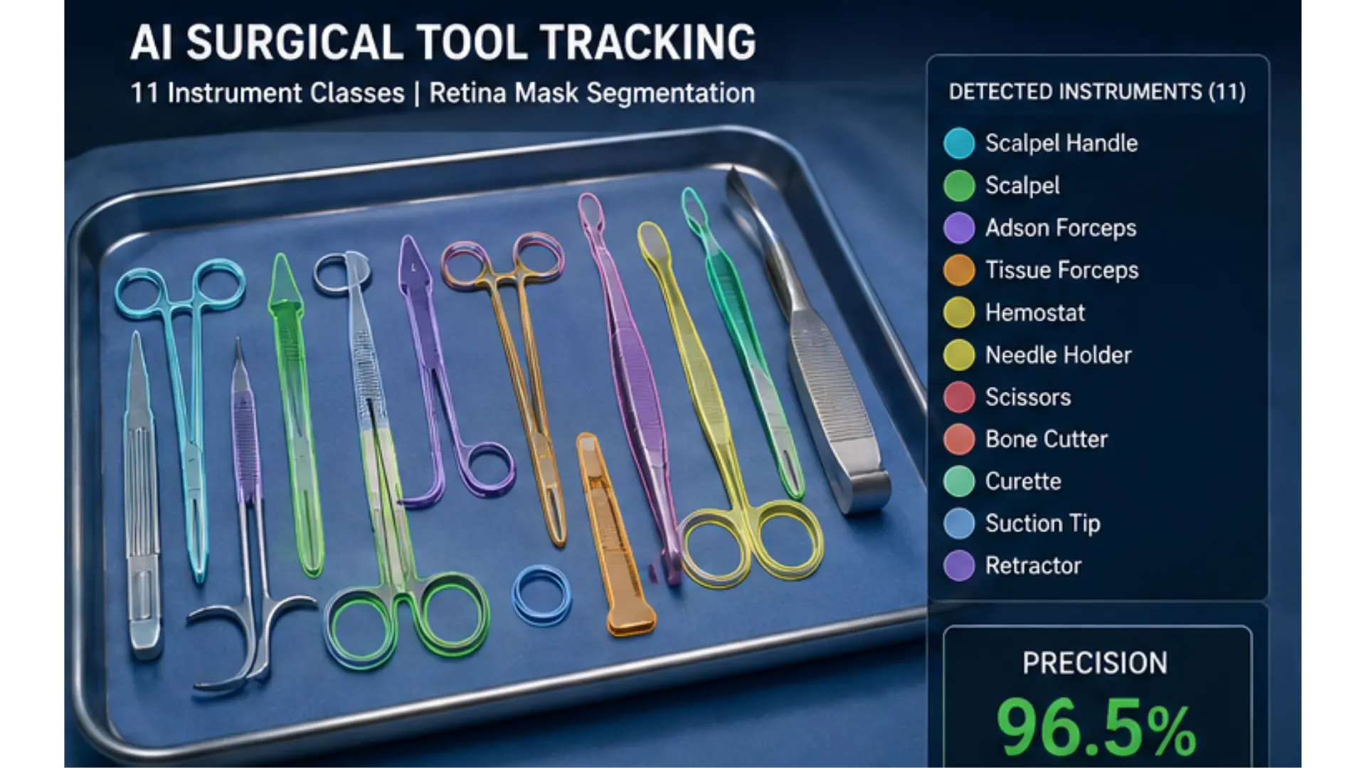
This project builds a computer vision system to automate that task. It uses a fine-tuned YOLOv11 Nano Segmentation model to detect, classify, and segment 11 distinct types of surgical instruments. Once a tool is detected, the system draws a precise visual mask over it. The same pipeline can also be used on high-resolution 4K video, where each frame is checked and the tools are highlighted.
The main goal is to improve safety and tracking. The system needs to know exactly what tools are on the table at all times. In this blog, we will look at how the system works, why YOLO 11 is useful for this task, how the masks are applied, and how the same idea can be used for real-time surgical video.
The Problem with Manual Tool Tracking
A standard operating room relies on humans to track tools. Nurses and technicians count the items before, during, and after a surgery. A manual system can follow a basic checklist, but this creates a major problem.
Human error happens. During long, stressful operations, the team is tired. There are dozens of tools on a tray. If the surgical team loses track of a tool, it can lead to severe consequences. Sometimes, items are accidentally left inside patients. This dangerous mistake is known as Retained Surgical Items, or RSI.
Checking the trays manually also takes a lot of time. If a surgeon has their hands inside a patient, they expect the right tool instantly. If a specific clamp is missing from the tray, the surgery stops. The team has to pause and find the missing item. This delay is not ideal when a patient is waiting. We need a system that can look at the tray and instantly know what is there.
How YOLO 11 Solves the Problem
The basic idea is simple. We give a camera system a clear view of the surgical tray. The system uses a YOLOv11 Nano architecture. The model looks at the image and identifies the tools. For example, it can instantly spot a Scalpel Handle or Forceps.
The model returns exact pixel boundaries for the regions it thinks match the tools. Each detection also has a confidence score. In this project, the model achieved a 96.5% precision score across 11 complex classes of metallic tools.
The Ultralytics Python package makes it easy to train the model and run predictions. The system converts its predictions into usable visual masks. In this project, a confidence threshold of 0.20 was used during video testing to account for the small training dataset. This allows the model to keep some weaker detections that may still be accurate.
Project Workflow
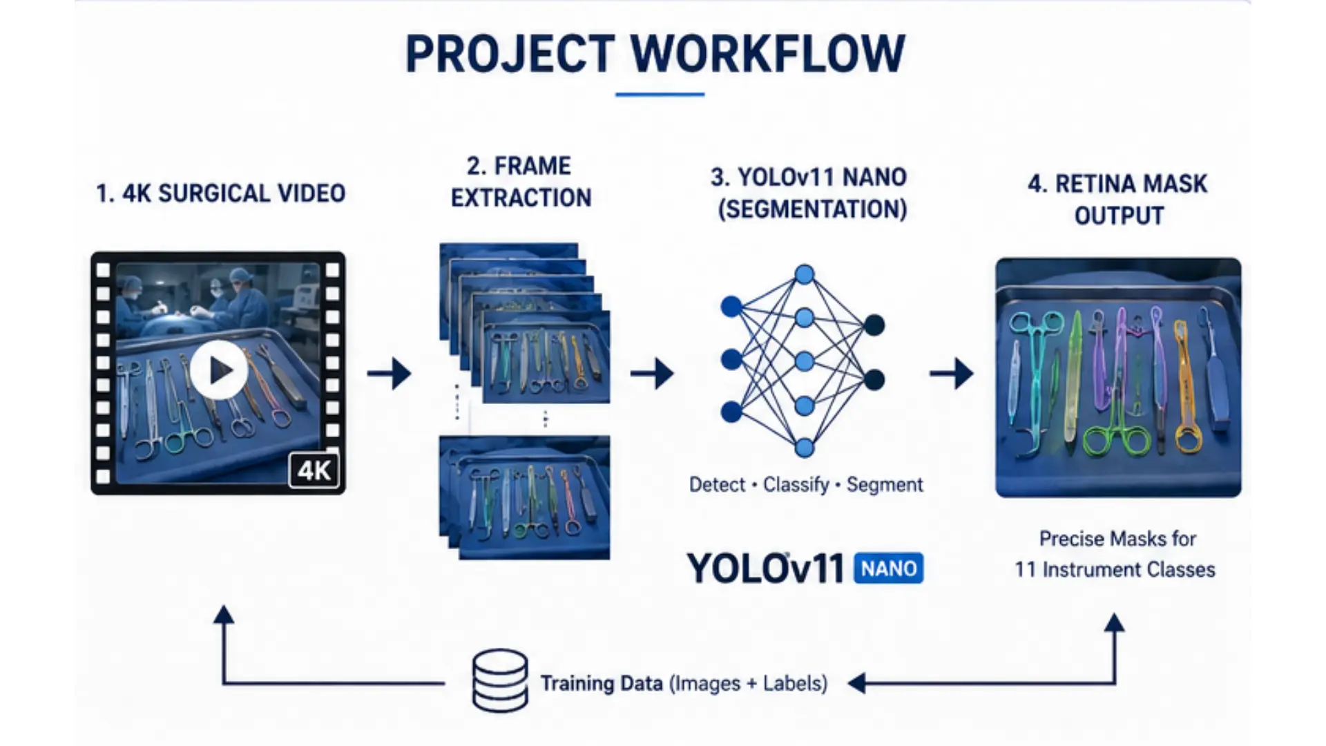
The complete system has four simple stages: frame extraction, format conversion, training, and video inference.
First, the system reads a high-resolution 4K surgical video. Random frames are extracted from this video to create a training dataset. Next, the labeled data is converted from a COCO format into the standard YOLO format. This creates specific folders for the training and validation images.
Project Workflow
The model then trains on these images. The model learns the shapes of the tools and returns a precise segmentation mask. Finally, the best trained model weights are loaded back into the software to run inference on the original 4K video.
The full process can be shown as: Video Frame Extraction → Format Conversion → YOLO11 Training → Output Inference.
This makes the system simple to understand and easy to extend.
Detecting Surgical Tools in Images
The first step was to test and train the system on individual images. Image testing is useful because it lets us teach the model before moving to a fast-moving video.
The video frames are extracted using a frame extractor tool. The images and labels are split into a training set and a validation set. The model then runs over these images and learns what the tools look like.
AI Surgical Tool Tracking
Each detection finds a general box area. More importantly, it finds the exact pixels inside that box that belong to the tool. This prepares the system to track the exact shapes of the instruments.
Why Use Segmentation Masks Instead of Bounding Boxes?
A simpler version of this project could just use basic bounding boxes. A bounding box draws a simple rectangle around the whole object. However, surgical tools are very thin and often sit very close together on the tray. A rectangle would capture a lot of the empty blue tray and overlap with other tools.
Precise segmentation is needed to know exactly where one tool ends and another begins. This project uses Retina Masks. Retina Masks ensure pixel-perfect boundaries on very thin metal instruments.
That produces a cleaner result. The mask follows the exact shape of the tool instead of covering a large rectangular area. Using a precise mask keeps the project highly accurate. A bounding box can create too much noise. A segmentation mask is much better for analyzing complex medical environments.
Improving the Training Quality
Training an AI usually requires thousands of images. However, this project used a micro-dataset strategy with a very small number of images. This makes it hard for the model to learn without just memorizing the pictures.
To fix this, the project used an advanced implementation of Backbone Freezing. Freezing protects the pre-trained knowledge the model already has. Heavy Contextual Augmentation was also applied. Techniques like Mosaic alter the images during training by mixing them together.
We also used HSV color adjustments to help the model handle lighting flickers in the video. This careful balancing allowed the model to successfully learn from an extremely limited dataset of around 100 instances.
Handling Video Inference
After the image training worked, the next step was to process a complete video. A video contains many individual frames. The system loads the best trained model weights and runs inference back on the original 4K video.
The model searches for tools, and the detected regions are highlighted with clean masks. During video inference, the standard bounding boxes and confidence numbers were turned off, leaving only the clean names and colorful masks.
The basic workflow is: Video → Frame → YOLO11 → Retina Mask → Output Frame.
Running deep learning on every frame of a 4K video can be slow. A large detection model needs significant computing power. However, this project strategically uses the YOLOv11 Nano architecture. The Nano version is very lightweight. It maintains incredibly high inference speeds on ultra-high-definition video without sacrificing precision.
Reflective Metallic Surfaces Are a Challenge
One important limitation appeared during testing. Not every tool is easy to see. Almost all surgical instruments are made of shiny metal. They strongly reflect the bright operating room lights.
Furthermore, many tools look visually similar to each other. A Bone Cutter and Forceps might look like the exact same shiny object to a basic camera. This means the model must be very smart to tell them apart.
Using Retina Masks helped solve this by keeping boundaries sharp. The model looks at the exact physical shape of the tool to figure out what it is, rather than getting confused by the bright, shiny reflections.
Real World Applications
A tool tracking system can be highly useful in many hospital workflows. An automated first pass can reduce the amount of manual counting needed.
The most important application is Operating Room Safety. Automated tracking prevents Retained Surgical Items by monitoring tool inventory before, during, and after surgery.
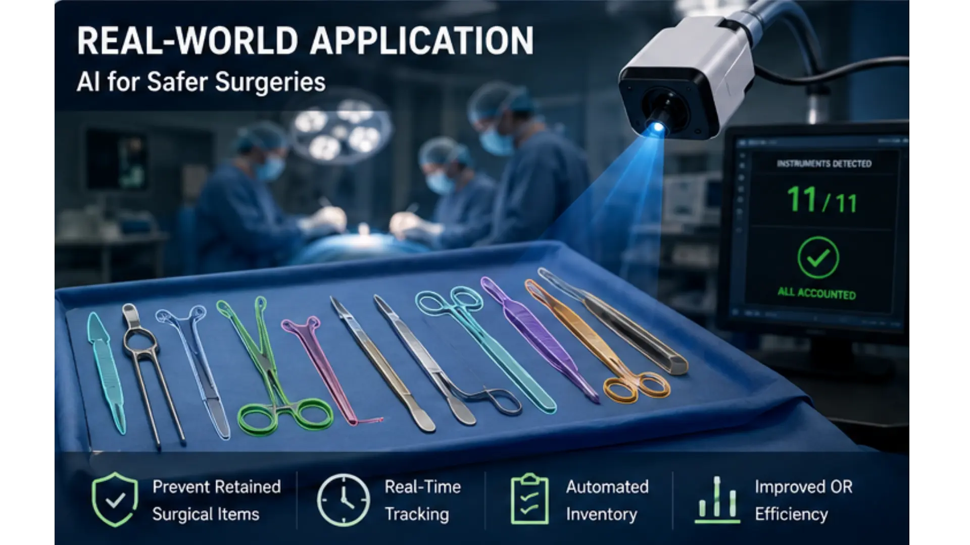
Real World Applications
The system also helps with Automated Inventory Management. It provides real-time tracking of sterile trays to ensure all required instruments are present before a procedure begins. This stops mid-surgery delays.
Another major use is Surgical Workflow Analysis. By tracking which tools are being used at what times, the system automatically documents the phases of a surgical procedure.
Finally, it can be used for Surgical Skill Assessment. Hospitals can analyze how often and how smoothly specific tools are handled to assess trainee surgeon performance.
Key Features of the System
Micro-Dataset Training Strategy The system uses an advanced implementation of Backbone Freezing and Heavy Contextual Augmentation. This allows it to successfully train an instance segmentation model on an extremely limited dataset.
High-Definition Segmentation The system utilizes Retina Masks. This ensures pixel-perfect boundaries on very thin, highly reflective metal instruments.
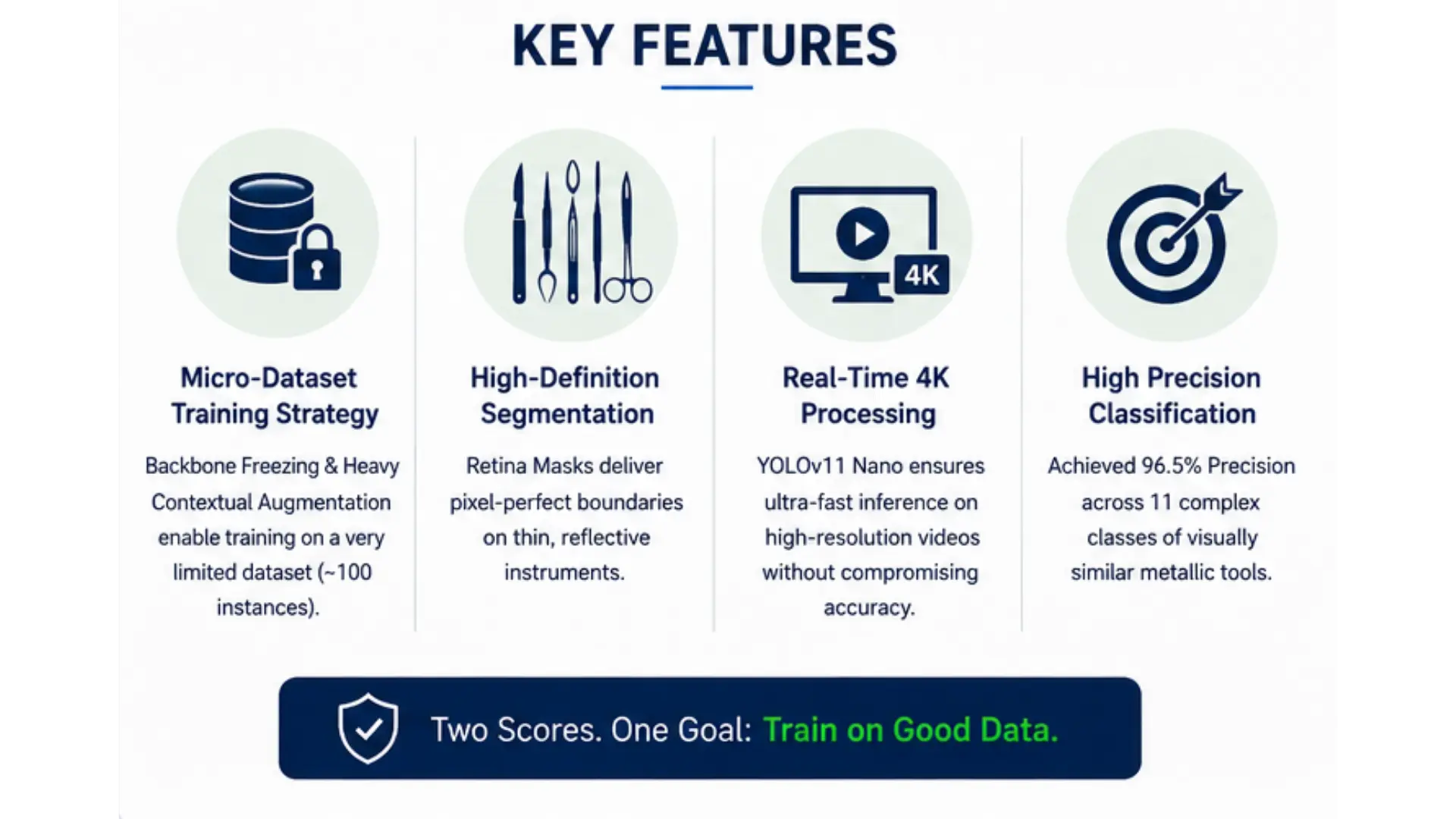
Key Features
Real-Time 4K Processing The same detection logic works on heavy video files. The strategic use of the YOLOv11 Nano architecture maintains incredibly high inference speeds on ultra-high-definition video without sacrificing precision.
High Precision Classification The model is highly accurate. It achieved 96.5% Precision across 11 complex classes of visually similar metallic tools.
Conclusion
This project shows how a modern computer vision model can be used for a highly critical medical task. Instead of relying entirely on human counting, we use an AI vision system to automatically spot, outline, and track 11 different surgical instruments.
The rest of the system is simple. The model finds the tool region, and the software draws a clean, precise mask over it. The same process is applied to 4K video frames to create an automated, real-time tracking system.
The current version proves that basic AI classification can solve massive real-world problems. We can use computer vision to verify trays and monitor surgeries instantly. This creates a strong base for a safer, more efficient, and less stressful operating room.
FAQs
How does AI prevent surgical tools from being left inside patients?
AI vision systems scan sterile surgical trays before, during, and after an operation to automatically track tool inventory. By verifying that every instrument is accounted for, the system prevents Retained Surgical Items (RSI).
Why use YOLO 11 for surgical tool tracking?
The YOLOv11 Nano architecture is lightweight, allowing it to maintain incredibly high inference speeds on ultra-high-definition 4K video without sacrificing precision. This ensures real-time tracking in fast-paced medical environments.
Why are segmentation masks better than bounding boxes for surgery?
Surgical tools are thin, highly reflective, and often placed close together. Pixel-perfect segmentation using Retina Masks precisely outlines the tool's exact shape, avoiding the overlap and noise caused by simple rectangular bounding boxes.

Simplify Your Data Annotation Workflow With Proven Strategies
.png)